
 MENU
MENU 

Students earn prizes for improving image processing techniques in EECS 556 (Winter 2014)
Two teams earned prizes in the graduate level course, EECS 556: Image Processing, thanks to the sponsorship of KLA-Tencor. The course, taught this past term by Prof. Jeff Fessler, covers the theory and application of digital image processing, which has applications in biomedical images, time-varying imagery, robotics, and optics. The KLA-Tencor judges in attendance this year were Eliezer Rosengaus and Jing Zhang.
First place
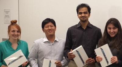
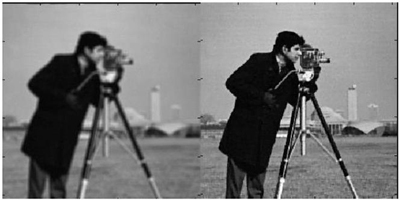
Unified Blind Method for Multi-Image Super-Resolution and Single/Multi-Image Blur Deconvolution
Rebecca Malinas, Yaohui Li, Abhishek Bafna, and Tatyana Dobreva
 Enlarge
“In many imaging applications,” explained the students, “an imaging system may produce low-resolution, blurry images. It is possible to improve the resolution of the output images through a process called super-resolution, in which we use multiple low-resolution images to recover a high-resolution image. However, the captured low-resolution images may also be blurry and noisy, which necessitates a deblurring step to recover a clear, high-resolution image. There is an additional problem with this, in that we do not know the blur kernel and so must simultaneously estimate the high-resolution image and the blur kernel. In our project, we investigated these processes and improved them through the use of color information in images. The use of color information greatly improved results.”
 Enlarge
 Enlarge
Second place
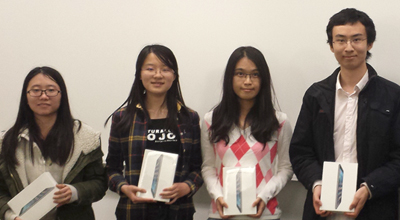
MRI bias field correction based on tissue labeling
Zhen Zeng, Lianli Liu, Jie Li, and Jiyang Chu
 Enlarge
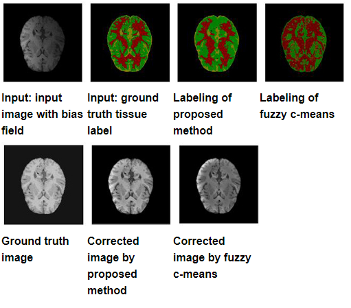 Enlarge
In magnetic resonance imaging (MRI), intensity nonuniformities, also known as the bias field, arise from a variety of factors. The human eye is able to taken into account this intensity inhomogeneity, allowing medical experts to analyze the MRI correctly. However, many intensity based image analysis algorithms are very sensitive to such intensity variation; thus correction of bias field is of great importance for accurate image analysis results. This project applied state-of-the-art segmentation techniques developed in computer vision to obtain better bias field estimation, and improved MRI images.